- Home
- Knee Joint Anatomy
Knee Joint Anatomy
Written By: Chloe Wilson, BSc(Hons) Physiotherapy
Reviewed by: KPE Medical Review Board
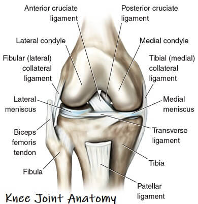
Knee joint anatomy involves looking at each of the different structures in and around the knee.
The knee joint is the largest and one of the most complex joints in the human body.
There are various muscles that control movement, ligaments that give stability, special cartilage to absorb pressure and various other structures to ensure smooth, pain-free movement.
Knee pain is a common problem that affects people at all ages. Many different things can go wrong with the knee. Understanding the anatomy of the knee and how the different parts fit together and function normally is the first step to understanding your knee problem and fixing it.
Functional Anatomy Of Knee Joint
The specific design of knee joint anatomy allows a number of functions:
- Supports the body in upright position without muscles having to work
- Helps in lowering and raising body e.g. sitting, climbing and squatting
- Allows rotation/twisting of the leg to place and position foot accurately
- Makes walking more efficient - just try walking without bending your knees and you'll see how much harder it is
- Acts with the ankle joint as a strong forward propeller of the body - particularly important when running
- Provides stability and proprioception of the leg
- Acts as a shock absorber
What Type of Joint Is The Knee?
The knee is what is known as a synovial hinge joint, but what does that mean?
- Synovial Joint: has a joint capsule which is like a sac surrounding the joint. The capsule contains synovial fluid which nourishes and lubricates the joint allowing it to move smoothly and painlessly - a bit like the oil in your car
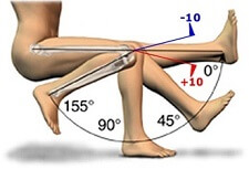
- Hinge Joint: typically allows motion in one plane, flexion and extension - think of it like a door hinge. The knee is the largest hinge joint in the body and is slightly unusual as it also allows a small amount of rotation
Knee Joint Anatomy & Function
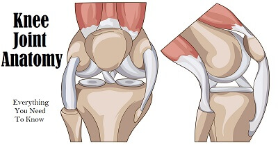
There are eight different components of lower leg and knee joint anatomy:
- Knee Bones
- Cartilage & Meniscus
- Leg & Knee Muscles
- Ligaments
- Tendons
- Bursa
- Joint Capsule
- Knee Cap
We start by looking at the features of each element of knee joint anatomy and then we will go on to look at the common problems that can develop with each of the different components of the anatomy of the knee.
1. Knee Bone Anatomy
The most basic component of knee joint anatomy are the bones which provide the structure to the knee.
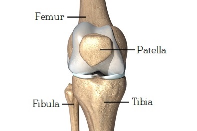
There are four knee bones that fit together to make two different knee joints:
- Femur: the thigh bone
- Patella: the kneecap
- Tibia: the main shin bone
- Fibula: the outer shin bone
The knee bones work together to support the entire body and transfer forces between the hip and foot, allowing the leg to move smoothly and efficiently.
Human knee joint anatomy actually comprises of two joints, the
- Tibiofemoral Joint: the main knee joint between the thigh and shin bones
- Patellofemoral Joint: the joint between the kneecap and thigh bone
You can find out all about the different bones and joints that make up the knee, how they function and what can go wrong in the knee bone anatomy section.
2. Knee Joint Cartilage Anatomy
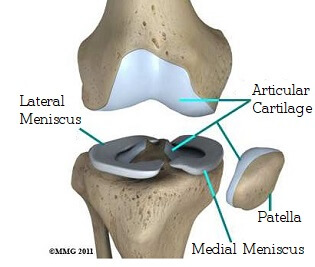
A really important part of knee joint anatomy is the cartilage. There are two different types of knee cartilage:
- Articular Cartilage: a thin layer of cartilage which lines the surfaces of the knee joints
- Knee Meniscus: a special extra thick layer of cartilage that sits on the top of the tibia
The knee meniscus is particularly important as it acts as a shock absorber to reduce the forces going through the bones and reduces friction, allowing the bones to move smoothly.
The back of the patella is also lined with cartilage, the thickest in the whole body due to the immense forces that go through the kneecap.
You can find out all about the different types of knee cartilage, how they work, and cartilage injuries and how to treat them in the knee joint cartilage anatomy section.
3. Knee & Leg Muscle Anatomy
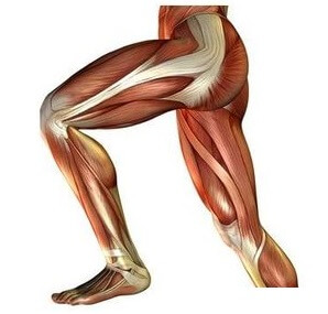
When looking at knee and leg muscle anatomy, the main knee muscles controlling the leg are the:
- Quadriceps: four muscles on
the front of the thigh which straighten the knee - rectus femoris, vastus medialis, vastus lateralis and vastus intermedius
- Hamstrings: three muscles on the back of the thigh which bend the knee - semimembranosus, semitendinosus and biceps femoris
- Glutes: three buttock muscles, very important in controlling the position of
the knee and how the forces transfer through the joint - gluteus maximus, gluteus medius and gluteus minimus
- Calves: two muscles on the back of the lower leg that control both the knee and ankle - gastrocnemius and soleus
Weakness and tightness in the leg muscles are common causes of knee pain. By stretching and strengthening the knee muscles, you can reduce the forces going through the knee joint, reducing pain and swelling, and improving function.
You can find out all about the different muscles in knee joint anatomy, how they work, how they can get damaged and how to strengthen and stretch them in the knee and leg muscle anatomy section.
4. Knee Ligament Anatomy
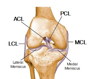
When thinking about knee joint anatomy in terms of stability, it is the knee ligaments that are most important as they are the main stabilising structures of the knee preventing excessive movements and instability. Ligaments are tough, fibrous connective tissues which link bone to bone, made of collagen.
There are two sets of knee ligaments:
- Cruciate Ligaments: anterior and posterior, ACL & PCL which sit inside the middle of the joint controlling forwards, backwards and twisting motion at the knee
- Collateral ligaments: medial and lateral, MCL & LCL that are found either side of the joint and control sideways stability of the knee
In the knee ligament anatomy section we look at each of the different ligament in-depth including how they work, what their role is, how they get damaged and how to make the best recovery from ligament injuries.
5. Knee Tendon Anatomy
Tendons are often overlooked as part of knee joint anatomy. They are they soft tissues found at the end of muscles which link the muscle to bone.
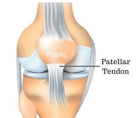
The main tendon found at the knee is the patellar tendon which links the quads muscles to the shin bone. The knee cap actually sits inside the patellar tendon.
Tendons are frequently damaged by overuse or excessive stretching resulting in tendonitis. The most common knee tendonitis problems are:
- Patellar Tendonitis: aka Jumpers Knee, just below the kneecap
- Quadricep Tendonitis: just above the kneecap
- Hamstring Tendonitis: at the back of the knee
6. Knee Bursa Anatomy
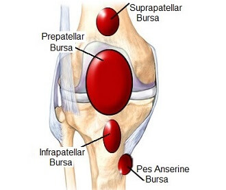
Now let's have a look at knee bursa anatomy. There are approximately fourteen bursa around the knee.
Bursa are small fluid filled sacs that reduce the friction between the bones and soft tissues to prevent inflammation.
If there is excessive friction on the bursa, usually due to muscle weakness, tightness or repetitive pressure, then the bursa gets inflamed, known as knee bursitis.
In the knee bursa anatomy section we look at each of the bursa, where they are, what they do, how they get injured and how to make a full recover from bursitis.
7. Knee Anatomy Joint Capsule
Another important part of knee anatomy is the joint capsule. This is like a bag that surrounds the joint containing synovial fluid to nourish and lubricate the knee allowing it to move smoothly and freely.
Any swelling that occurs in the joint is contained inside the capsule, which is why injuries can cause the knee to "balloon". If the joint capsule is damaged, swelling is no longer confined to the joint so tends to actually be less obvious.
You can find out more in the knee swelling section.
8. Knee Anatomy Patella
When looking at the anterior aspect of knee joint anatomy, you will find the patella (kneecap). The patella is a small, triangular bone which sits in the quadriceps muscle at the front of the knee.
The kneecap is lined with cartilage which is actually the thickest cartilage in the whole body due to the massive forces that go through it e.g. the force going through the kneecap when coming downstairs is 3.5x body weight.
Knee cap pain is one of the most common problems associated with the knee.
Common Knee Injuries
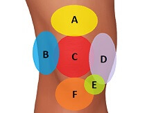
There are lots of things that can go wrong with knee joint anatomy, either from an injury or from gradual wear and tear:
- Meniscus Injuries
- Knee Ligament Injuries
- Kneecap Injuries
- Knee Bursitis
- Tendonitis
- Muscle Tears
- Bone Spurs In Knee
Knee injuries typically result in a combination of pain, instability and reduced knee range of motion. To find out more, check out the sections on knee injuries and common problems.
Alternatively, if you have a knee problem and want help working out what is wrong, visit the knee pain diagnosis section.
You can find out more in-depth information on the different aspects of anatomy of the knee:
There you will find everything you need to know about knee joint anatomy.
Related Articles
Diagnosis Chart
March 25, 2025
Knee Pain On Stairs
January 16, 2023
Page Last Updated: March 25th, 2025
Next Review Due: March 25th, 2027
